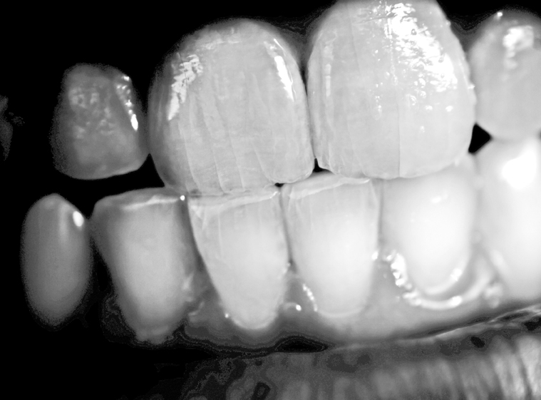
For years we have searched for answers regarding why some of our patients clench and/or grind their teeth. We have blamed this so called “parafunctional activity” on everything from premature occlusal contacts, dysfunctional mandibular position (too far forward, too far backward, too much vertical, too little vertical, in “CR,” not in “CR,”) and of course, emotional and psychological stress. We often see the results of bruxism in children and hear from the parents of the violent and loud nature of the grinding. We have provided every type of plastic to place between the teeth of patients to “control” their parafunctional behavior, or at least to try to protect their teeth and periodontium from damage. But often our attempts to control the behavior seem futile, and the patient continues to fight whatever piece of plastic we place in their mouth, or worse yet, destroy their teeth and restorations.
Speaking of restorations, have you noticed that patients with significant anterior wear tend to break whatever restorations you place, be it composite, veneers, or even full crowns? Why are these patients grinding on their front teeth to begin with? If you ask the patient if they are aware of grinding on their front teeth they will almost always tell you that they are not aware, and likely the patient is indeed not grinding anteriorly during the day.
Then you have the patient with a beautiful occlusion.Almost too perfect. Then you notice wear facets on the posterior teeth. This patient is more likely to have fractured posterior restorations, and are the patients that “chew through” their nightguards. They are also more likely to suffer with morning headaches and various “TMJ” disorders. They too are usually unaware of spending a lot of time during the day clenching. They may acknowledge clenching from time to time, but not enough to cause the wear and effects that you see.
So why are these patients grinding and/or clenching their teeth while they sleep?
Over the past 30 years, a few practitioners and researchers have looked at the connection between the airway and temporomandibular disorders, particularly from a growth and development standpoint. In the last 20 years there have been more and more studies looking at exactly what is happening during bruxism events. These parafunctional activities have become better defined and described, but the “why” still remained.
However, recent studies indicate that in many cases so called “parafunctional activity” may actually be an attempt by the central and autonomic nervous systems to protect the organism (1-4). As dentists have become more involved in the treatment of obstructive sleep apnea, we are learning how very important the airway is. We’ve always recognized the “ABC’s” and know that “airway and breathing” are first and foremost, because without a proper airway we can’t breathe, and if we can’t breathe, we’re dead.
One study that should keep you awake at night (pun intended) is a pilot study which showed that five of ten subjects with sleep apnea had their sleep apnea WORSEN when using a flat plane nightguard, just like the ones most dentists use to “protect” their patients from bruxism. The authors state in their conclusion:
This open study suggested that the use of an occlusal splint is associated with a risk of aggravation of respiratory disturbances. It may therefore be relevant for clinicians to question patients about snoring and sleep apnea when recommending an occlusal splint.
That’s exactly what I’m saying.
I believe what is happening is that the patient’s brain is attempting to maintain a patent airway. This is done by either grinding anteriorly (think “head tilt, chin lift”, or the patient bringing their chin forward), by clenching the teeth to keep the jaw from falling back, or by some combination of the two. If this hypothesis is true, then we should see less bruxism in patients with sleep apnea when the sleep apnea is effectively treated.
And that’s exactly what we do see.
A case study reported that a patient with severe sleep apnea who also exhibited 61 bruxism events on the baseline study had “complete eradication of the tooth grinding events” when continuous positive airway pressure (CPAP) was introduced. The authors state that “when sleep bruxism is related to apnea/hypopneas, the successful treatment of these breathing abnormalities may eliminate bruxism during sleep.” (6)
This is also seen in children. When children are diagnosed with sleep apnea the typical first line therapy is adenotonsillectomy. Several studies have shown a reduction of bruxism events after removal of the tonsils. (7-8) Most dentists believe that children grow out of the bruxism activity, and that may be the case for some. However, I believe that a lot of children don’t grow out of it, it just takes longer for the very hard enamel of the permanent teeth to show the wear patterns, as opposed to the relatively rapid breakdown of the significantly softer and thinner enamel of the primary dentition.
The good news is that when bruxism is related to airway issues, appropriate therapy, including use of mandibular advancement devices in adults, can reduce or even eliminate the bruxism. Knowing this can help you protect your patient’s teeth, protect your restorations (saving you thousands in remakes,) and literally protect your patients’ lives.
In a furture article I will discuss what to look for when evaluating your patients for possible sleep apnea-related bruxism, how to refer for medical evaluation when indicated, and how to manage and treat these patients, both from a restorative perspective as well as helping them with their sleep apnea.
Dr. Jamison Spencer is the director of the Craniofacial Pain Center of Idaho in Boise and the Craniofacial Pain Center of Colorado in Denver. Dr. Spencer is the Past President of the American Academy of Craniofacial Pain (AACP), a Diplomate of the American Board of Craniofacial Pain, a Diplomate of the American Board of Dental Sleep Medicine and has a Masters with a certificate in Craniofacial Pain from Tufts University. He teaches head and neck anatomy at Boise State University and is adjunct faculty at the Tufts Craniofacial Pain Center in both the craniofacial pain residency and dental sleep medicine programs. Dr. Spencer lectures nationally and internationally on TMD, dental sleep medicine and head and neck anatomy and is faculty of the AACP’s Institute and the AACP/Tufts Dental Sleep Medicine program.
Dr. Spencer lives in Boise Idaho with his wife of 21 years Jennifer and their 6 children.
1. A significant increase in breathing amplitude precedes sleep bruxism. Khoury S, Rouleau GA, Rompré PH, Mayer P, Montplaisir JY, Lavigne GJ. Chest. 2008 Aug;134(2):332-7. Epub 2008 May 19.
2. Genesis of sleep bruxism: motor and autonomic-cardiac interactions. Lavigne GJ, Huynh N, Kato T, Okura K, Adachi K, Yao D, Sessle B. Arch Oral Biol. 2007 Apr;52(4):381-4. Epub 2007 Feb 20.
3. Association between sleep bruxism, swallowing-related laryngeal movement, and sleep positions. Miyawaki S, Lavigne GJ, Pierre M, Guitard F, Montplaisir JY, Kato T. Sleep. 2003 Jun 15;26(4):461-5.
4. Effect of sleep position on sleep apnea and parafunctional activity. Phillips BA, Okeson J, Paesani D, Gilmore R. Chest. 1986 Sep;90(3):424-9.
5. Aggravation of respiratory disturbances by the use of an occlusal splint in apneic patients: a pilot study. Gagnon Y, Mayer P, Morisson F, Rompré PH, Lavigne GJ. Int J Prosthodont. 2004 Jul-Aug;17(4):447-53.
6. Sleep bruxism related to obstructive sleep apnea: the effect of continuous positive airway pressure. Oksenberg A, Arons E. Sleep Med. 2002 Nov;3(6):513-5.
7. Improvement of bruxism after T&A surgery. DiFrancesco RC, Junqueira PA, Trezza PM, de Faria ME, Frizzarini R, Zerati FE. Int J Pediatr Otorhinolaryngol. 2004 Apr;68(4):441-5.
8. Bruxism and adenotonsillectomy. Eftekharian A, Raad N, Gholami-Ghasri N. Int J Pediatr Otorhinolaryngol. 2008 Apr;72(4):509-11. Epub 2008 Feb 20.


